| 2020 №03 (07) |
DOI of Article 10.37434/sem2020.03.08 |
2020 №03 (01) |

Сучасна електрометалургія, 2020, #3, 54-61 pages
Електронно-променевий синтез наночастинок оксиду заліза та їх біологічна активність
С.Є. Литвин1, Ю.А. Курапов1, О.М. Важнича2, Я.А. Стельмах1, С.М. Романенко1, О.І. Оранська3
1ІЕЗ ім. Є.О. Патона НАН України. 03150. м. Київ, вул. Казимира Малевича, 11. E-mail: office@paton.kiev.ua
2Українська медична стоматологічна академія. 36011, м. Полтава, вул. Шевченка, 23. E-mail: vazhnichaya@ukr.net
3Інститут хімії поверхні ім. О.О. Чуйка НАН України. 03164, м. Київ, вул. Генерала Наумова, 17. E-mail: el.oranska@gmail.com
Реферат
Наведено результати дослідження структури пористих конденсатів композиції Fe–NaCl, хімічного, фазового складів і розміру наночастинок, отриманих фізичним синтезом з парової фази з використанням методу EB PVD. При швидкому вилученні з вакууму наночастинки заліза окислюються на повітрі до магнетиту. У початковому стані вони мають велику сорбційну здатність по відношенню до кисню і вологи, тому при подальшому нагріванні на повітрі відбувається зниження маси пористого конденсату аж до температури 650 °С за рахунок десорбції фізично сорбованої вологи. Фізично адсорбований кисень приймає участь в доокисленні Fe3O4 до Fe2O3. Збільшення температури конденсації супроводжується зростанням розміру наночастинок, в результаті чого значно скорочується їх сумарна площа поверхні та сорбційна здатність. Навіть без стабілізації такі наночастинки, досліджувані у вигляді водного колоїду, виготовленого ex tempore, мають в експерименті на тваринах характерну протианемічну дію, що може бути використано у медицині. Бібліогр. 41, табл. 2, рис. 5.
Ключові слова: EB PVD; наночастинки оксиду заліза; сорбція; фазовий склад; колоїдні системи; антианемічний ефект
Received 01.11.2019
Список літератури
1. Willard, M.A., Kurihara, L.K., Carpenter, E.E. et al. (2004) Chemically prepared magnetic nanoparticles. Int. Materials Reviews, 49, 125–170. doi.org/10.1179/095066004225021882.2. Cabuil, V. Magnetic nanoparticles (2008) Dekker encyclopedia of nanoscience and nanotechnology, V. 3, CRC Press, Taylor and Francis Group, Boca Raton, Fl; 1985–2000. https://www.amazon.com/Dekker-Encyclopedia-Nanoscience-Nanotechnology-3/dp/0824750497
3. Revia, R.A., Zhang, M. (2016) Magnetite nanoparticles for cancer diagnosis, treatment, and treatment monitoring: recent advances. Materials Today, 19(3), 157–168. https://dx.doi.org/10.1016%2Fj.mattod.2015.08.022
4. Nikolaev, V.I., Shipilin, A.M., Zakharova, I.N. (2001) On estimating nanoparticle size with the help of the Mössbauer effect. Physics of the Solid State, 43, 1515–1517. doi.org/10.1134/1.1395093
5. Thach, C.V., Hai, N.H., Chau, N. (2008) Size controlled magnetite nanoparticles and their drug loading ability. J. of the Korean Phys. Soc., 52, 1332–1335. doi.org/10.3938/jkps.52.1332
6. Shpak, A.P., Gorbyk, P.P. (2009) Nanomaterials and supramolecular structures: Physics, chemistry, and applications, Dordrecht, London New York, Springer. http://www.springer. com/gp/book/9789048123087
7. Yu, J., Yang, C., Li, J. et al. (2014) Multifunctional Fe5C2 nanoparticles: a targeted theranostic platform for magnetic resonance imaging and photoacoustic tomography-guided photothermal therapy. Advanced Materials, 26(24), 4114– 4120. doi.org/10.1002/adma.201305811
8. Ju, Y., Zhang, H., Yu, J. et al. (2017) Monodisperse Au–Fe2C Janus nanoparticles: An attractive multifunctional material for triple-modal imaging-guided tumor photothermal therapy. ACS Nano, 11(9), 9239–9248. doi.org/10.1021/acsnano. 7b04461
9. Kopanja, L., Kralj, S., Zunic, D. et al. (2016) Core–shell superparamagnetic iron oxide nanoparticle (SPION) clusters: TEM micrograph analysis, particle design and shape analysis. Ceramics Int., 42, 10976–10984. doi.org/10.1016/j.ceramint. 2016.03.235
10. Tadić, M., Kralj, S., Jagodic, M. et al. (2014) Magnetic properties of novel superparamagnetic iron oxide nanoclusters and their peculiarity under annealing treatment. Applied Surface Sci., 322, 255–264. doi.org/10.1016/j.apsusc.2014.09.181
11. Wang, X., Zhang, P., Wang, W. et al. (2016) Magnetic N-enriched Fe3C/graphitic carbon instead of Pt as an electrocatalyst for the oxygen reduction reaction. Chem. Eur. J., 22, 4863–4869. doi.org/10.1002/chem.201505138
12. Elmore, W.C. (1938) Ferromagnetic colloid for studying magnetic structures. Phys. Rev., 54, 309–310. doi.org/10.1103/PhysRev.54.309
13. Klabunde, K., Sergeev, G.B. (2013) Nanochemistry. Elsevier, 2nd ed. https://www.elsevier.com/books/nanochemistry/klabunde/ 978-0-444-59397-9
14. Cushing, B.L., Kolesnichenko, V.L., O’Connor, C.J. (2004) Recent advances in the liquid-phase syntheses of inorganic nanoparticles. Chem. Rev., 104, 3893–3946. doi.org/10.1021/cr030027b
15. Burda, C., Chen, X., Narayanan, R., El-Sayed, M.A. (2005) Chemistry and properties of nanocrystals of different shapes. Ibid., 105(4), 1025–1102. doi.org/10.1021/cr030063a
16. Kopanja, L., Milošević, I., Panjan, M. et al. (2016) Sol–gel combustion synthesis, particle shape analysis and magnetic properties of hematite (α-Fe2O3) nanoparticles embedded in an amorphous silica matrix. Applied Surface Sci., 362, 380– 386. doi.org/10.1016/j.apsusc.2015.11.238
17. Tadić, M., Kusigerski, V., Marković, D. et al. (2012) Highly crystalline superparamagnetic iron oxide nanoparticles (SPION) in a silica matrix. J. of Alloys and Compounds, 525, 28–33. doi.org/10.1016/j.jallcom.2012.02.056
18. Billas, I.M.L., Chatelain, A., de Heer, W.A. (1997) Magnetism of Fe, Co and Ni clusters in molecular beams. J. Magn. Magn. Mater., 168(1–2), 64–84. doi.org/10.1016/S0304-8853(96)00694-4
19. Billas, I.M.L., Chatelain, A., de Heer, W.A. (1996) Magnetism in transition-metal clusters from the atom to the bulk. Surface Review and Letters, 3(1), 429–434. doi.org/10.1142/S0218625X96000772
20. Gubin, S.P., Koksharov, Yu.A., Khomutov, G.B., Yurkov, G.Yu. (2005) Magnetic nanoparticles: Preparation, structure and properties. Russ. Chem. Rev., 74(6), 489–520. doi.org/10.1070/RC2005v074n06ABEH000897 [in Russian].
21. Roca, A.G., Costo, R., Rebolledo, A.F. et al. (2009) Progress in the preparation of magnetic nanoparticles in biomedicine. J. of Physics D: Applied Physics, 42(22), 224002. doi.org/10.1088/0022-3727/42/22/224002
22. Movchan, B.A., Kurapov, Yu.A., Didikin, G.G. et al. (2011) Regulation of composition and structure of nanoparticles of Fe-O system in electron beam evaporation of Fe3O4. Poroshk. Metallurgiya, 478(3–4), 56–63. doi.org/10.1007/s11106-011-9314-0 [in Russian].
23. Kurapov, Yu.A., Krushinskaya, L.A., Litvin, S.E. et al. (2014) Producing of silver nanoparticles and their thermal stability in Ag-O system. Ibid., 496(3–4), 95–102. doi.org/10.1007/s11106-014-9604-4 [in Russian].
24. Kurapov, Yu.A., Litvin, S.E., Romanenko, S.M. (2013) Structure and thermal stability of Ti–NaCl condensates deposited from the vapour phase in vacuum. Nanostructured Materials Sci., 1, 55–62. http://www.materials.kiev.ua/science/edition_view.jsp?id=2 [in Russian]. 25. Kurapov, Yu.A., Litvin, S.E., Didikin, G.G., Romanenko, S.M. (2011) Structure of two-phase condensates Cu–NaCl deposited from vapor phase in vacuum. Sovrem. Elektrometall., 2, 19–22. https://patonpublishinghouse.com/eng/journals/ sem/2011/02/05 [in Russian]. 26. Kurapov, Yu.A., Romanenko, S.M., Didikin, G.G., Oranskaya, E.I. (2017) Controllable synthesis of iron oxide nanoparticles in porous NaCl matrix. Materials Research Express, 4(3), 035031. doi.org/10.1088/2053-1591/4/3/035031 27. Kovinsky, I.S., Krushinskaya, L.A., Movchan, B.A. (2011) Structure and some properties of sodium chloride condensates produced by electron beam evaporation with next deposition in vacuum. Sovrem. Elektrometall., 1 , 42–46. https:// patonpublishinghouse.com/eng/journals/sem/2011/01/08 [in Russian].
28. Chekman, I.S., Ul’berh, Z.R., Malanchuk, V.O. et al. (2012) Nanoscience, nanobiology, nanofarmation. Kyiv, Polihraf plyus. https://www.twirpx.com/file/115774/
29. Laurent, S., Forge, D., Port, M. et al. (2008) Magnetic iron oxide nanoparticles: Synthesis, stabilization, vectorization, physicochemical characterizations, and biological applications. Chem. Rev., 108(6), 2064–2110. doi.org/10.1021/cr068445e
30. Santhosh, P.B., Ulrih, N.P. (2013) Multifunctional superparamagnetic iron oxide nanoparticles: Promising tools in cancer theranostics. Cancer Lett., 336(1), 8–17. doi.org/10.1016/j.canlet.2013.04.032
31. Jin, R., Lin, B., Li, D., Ai, H. (2014) Superparamagnetic iron oxide nanoparticles for MR imaging and therapy: Design considerations and clinical applications. Curr. Opin. Pharmacol., 18, 18–27. doi.org/10.1016/j.coph.2014.08.002
32. Wang, Y.X.J. (2011) Superparamagnetic iron oxide based MRI contrast agents: Current status of clinical application. Quant. Imaging Med. Surg., 1(1), 35–40. doi.org/10.3978%2Fj.issn.2223-4292.2011.08.03
33. Rosner, M.H., Auerbach, M. (2011) Ferumoxytol for the treatment of iron deficiency. Expert Rev. Hematol., 4(4), 399–406. doi.org/10.1586/ehm.11.31
34. Roohi, F., Lohrke, J., Ide, A. et al. (2012) Studying the effect of particle size and coating type on the blood kinetics of superparamagnetic iron oxide nanoparticles. Int. J. Nanomedicine, 7, 4447–4458. doi.org/10.2147%2FIJN.S33120
35. Ni, F., Jiang, L., Yang, R. et al. (2012) Effects of PEG length and iron oxide nanoparticles size on reduced protein adsorption and non-specific uptake by macrophage cells. J. Nanosci. Nanotechnol., 12(3), 2094–2100. doi.org/10.1166/jnn.2012.5753
36. Briley-Saebo, K.C., Johansson, L.O., Hustvedt, S.O. et al. (2006) Clearance of iron oxide particles in rat liver: effect of hydrated particle size and coating material on liver metabolism. Invest. Radiol., 41(7), 560–571. doi.org/10.1097/01.rli.0000221321.90261.09
37. Paton, B.E., Movchan, B.O., Kurapov, Yu.A., Yakovchuk, K.Yu. (2010) Method of producing of metal-oxygen system nanoparticles with specified composition by electron beam evaporation and condensation in vacuum. Ukraine, Pat. 92556. http://base.ukrpatent.org/searchINV/search.php?action=viewdetails&IdClaim=151646 [in Ukrainian].
38. Lebedev, A.D., Levchuk, Y.N., Lomakin, A.V. (1987) Clearance of iron oxide particles in rat liver: Effect of hydrated particle size and coating material on liver metabolism. Ed. by V.A. Noskin. In: Laser correlation spectroscopy in biology. Kiev, Naukova Dumka. https://search.rsl.ru/ru/record/01001388286 [in Russian].
39. Merkus, H.G. (2009) Particle size measurements. Fundamentals, practice, quality, Springer. http://www.springer.com/gp/book/9781402090158
40. Vazhnycha, O.M., Moklyak, E.B., Movchan, B.O., Kurapov, Yu.A. Method of treatment of acute hemorrhage using the nanoparticles of iron oxide. Ukraine, Pat. 103401 http://uapatents.com/6-103401-sposib-likuvannya-gostro-krovovtrati-za-dopomogoyu-nanochastinok-oksidu-zaliza-ii-iii.html [in Ukrainian].
41. Litvin, S.E.,Kurapov, Yu.A., Vazhnichaya, E.M. et al. (2019) Synthesis of iron oxide nanoparticles by method of electron beam deposition in vacuum. In: Proc. of 9th Int. Conf. on Beam Technologies in Welding and Materials Processing. Ed. by I.V. Krivtsun. IAW, 63–70. https://patonpublishinghouse.com/proceedings/ltwmp2019.pdf
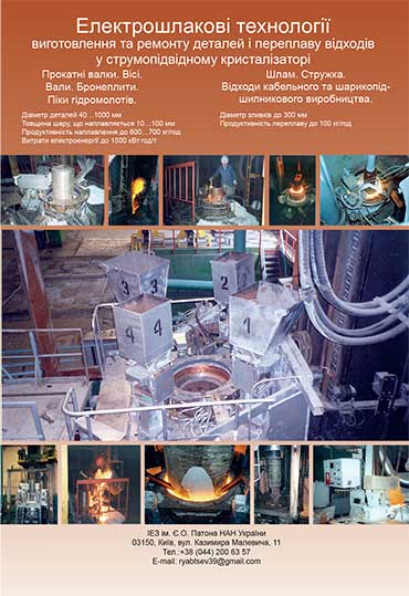
Реклама в цьому номері:
Вартість передплати/замовлення на журнали або окремі статті
| журнал/валюта | річний комплект друкований |
1 прим. друкований |
1 прим. електронний |
одна стаття (pdf) |
| AS/UAH | 1800 грн. | 300 грн. | 300 грн. | 150 грн. |
| AS/USD | 192 $ | 32 $ | 26 $ | 13 $ |
| AS/EUR | 180 € | 30 € | 25 € | 12 € |
| TPWJ/UAH | 7200 грн. | 600 грн. | 600 грн. | 280 грн. |
| TPWJ/USD | 384 $ | 32 $ | 26 $ | 13 $ |
| TPWJ/EUR | 348 € | 29 € | 24 € | 12 € |
| SEM/UAH | 1200 грн. | 300 грн. | 300 грн. | 150 грн. |
| SEM/USD | 128 $ | 32 $ | 26 $ | 13 $ |
| SEM/EUR | 120 € | 30 € | 25 € | 12 € |
| TDNK/UAH | 1200 грн. | 300 грн. | 300 грн. | 150 грн. |
| TDNK/USD | 128 $ | 32 $ | 26 $ | 13 $ |
| TDNK/EUR | 120 € | 30 € | 25 € | 12 € |
AS = «Автоматичне зварювання» - 6 накладів на рік;
TPWJ = «PATON WELDING JOURNAL» - 12 накладів на рік;
SEM = «Сучасна електрометалургія» - 4 наклада на рік;
TDNK = «Технічна діагностика та неруйнівний контроль» - 4 наклада на рік.


